|
|
Sialolithiasis
Stone in Wharton's Duct
- Most common disease of salivary glands
- Twice as common in males as females
- 80-95% occur in submandibular gland or duct
- Stones are most common cause of acute and chronic infection of salivary glands
- 80% of submandibular stones are opaque; 60% of parotid are opaque
- Consist of mainly calcium phosphate
- Not associated with systemic calcium abnormalities
- Very unusual for patients to have a combination of radiopaque and non-opaque stones
- Signs and symptoms
- Pain and swelling of involved gland
- Sialolithiasis causes pain and swelling of the involved salivary gland by obstructing the food-related flow of salivary secretions
- Calculi may cause stasis of saliva facilitating bacterial ascent into the gland and subsequent infection
- Some may be asymptomatic
- Imaging
- Plain radiography
- Opaque stone in course of Wharton’s (submandibular) or Stensen’s (parotid) ducts
- CT
- Stone in duct
- Ductal dilatation
- MR
- Sialography is contraindicated in acute infection or in a patient with a significant contrast allergy
- Treatment
- Conservative
- Surgical removal
- Lithotripsy
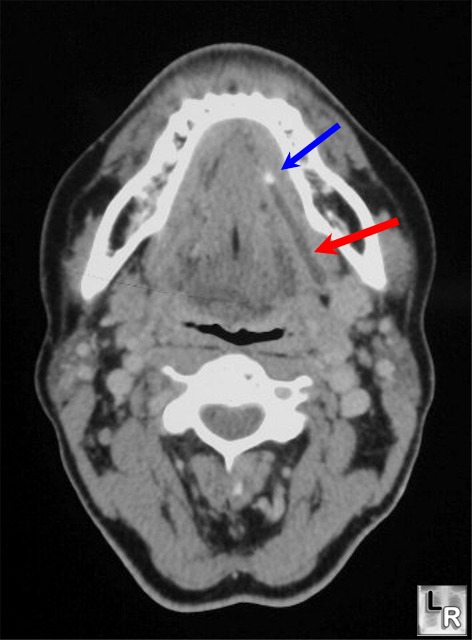
Sialolithiasis. Contrast-enhanced CT of the neck demonstrates
a stone (blue
arrow) in the submandibular region of a dilated
Wharton's Duct
(red arrow).
|
|
|
