|
|
Porcelain Gallbladder
- Calcification of gallbladder wall
- So named because of its gross appearance and its
similarity to porcelain
- Incidence:
- Less than 1% of cholecystectomy patients
- F:M 5:1
- Histology
- Flakes of dystrophic calcium within chronically
inflamed and fibrotic muscular wall
- Wall is thickened and gallbladder is contracted
- Associated with gallstones in 90%
- Cystic duct is always obstructed
- 80% of patients with carcinoma of gallbladder have
stones
- Minimal symptoms
- Imaging findings
- Curvilinear calcifications in segment of the wall
or entire wall
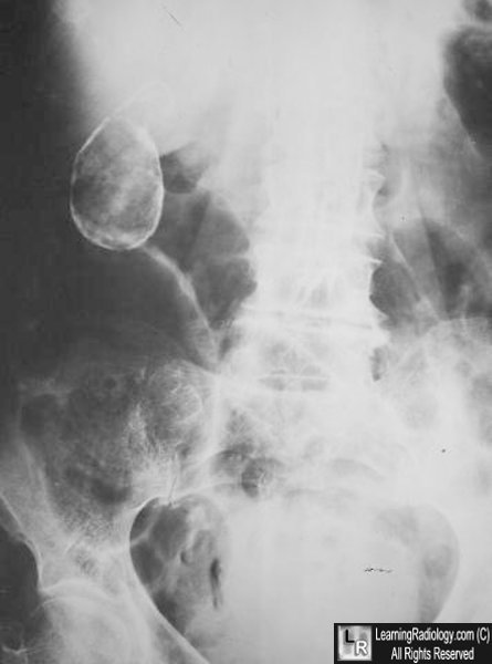
Porcelain Gallbladder. Plain film of abdomen shows a curvilinear calcification
in the
right upper quadrant which corresponds to the location of the gallbladder
- Highly echogenic shadowing curvilinear structure in
GB fossa
- DDx: stone-filled contracted GB
- Echogenic GB wall with little acoustic shadowing
- DDx: emphysematous cholecystitis
- Scattered irregular clumps of echoes with posterior
acoustic shadowing
- Imaging pitfall
- Contracted gallbladder with calcified wall can be
mistaken for a gallstone
- Complication
- 20-30% develop carcinoma of gallbladder
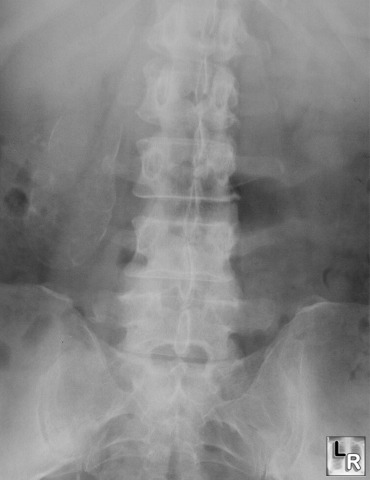
Porcelain Gallbladder. Plain film of abdomen shows a curvilinear
calcification in the
right upper quadrant which corresponds to the location of the
gallbladder
|
|
|
