|
|
Malignant Mesothelioma
- Most common primary neoplasm of pleura
- Prevalence
- 2,000-3,000 cases/year in US
- Etiology
- Asbestos exposure
- Zeolite (non-asbestos mineral fiber)
- Chronic inflammation (TB, empyema)
- Radiation
- Peak age
- Histology
- Epithelioid (60%)
- Sarcomatoid (15%)
- Biphasic (25%)
- Intracellular asbestos fibers in 25%
- Carcinogenic potential: crocidolite > amosite > chrysotile > actinolite, anthophyllite, tremolite
- Occupational exposure of asbestos found in only
40-80% of all cases
- 5-10% of asbestos workers will develop
mesothelioma (risk factor of 30X compared with general population)
- No relation to duration/degree of exposure to
asbestos or smoking history
- Latency period
- 20-45 years
- Earlier than asbestosis
- Later than asbestos-related lung cancer
- Pathology
- Multiple tumor masses involving predominantly
the parietal pleura and to a lesser degree the visceral pleura
- Progresses to thick sheet-like / confluent masses resulting in lung encasement
- Associated with
- Peritoneal mesothelioma
- Hypertrophic osteoarthropathy (10%)
- Staging (Boutin modification of Butchart staging)
- IA confined to ipsilateral parietal /
diaphragmatic pleura
- IB+ visceral pleura, lung , pericardium
- II invasion of chest wall / mediastinum
(esophagus, heart, contralateral pleura) or metastases to thoracic
lymph nodes
- III penetration of diaphragm with peritoneal
involvement or metastases to extrathoracic lymph nodes
- IV distant hematogenous metastases
- Stage at presentation
- II in 50%
- III in 28%
- I in 18%
- IV in 4%
- Clinical signs and symptoms
- Non-pleuritic (56%) /
pleuritic chest pain (6%)
- Dyspnea (53%)
- Fever + chills + sweats (30%)
- Weakness, fatigue, malaise (30%)
- Cough (24%)
- Weight loss (22%)
- Anorexia (10%)
- Expectoration of asbestos bodies (= fusiform
segmented rod-like structures =
iron-protein deposition on asbestos fibers [a subset of ferruginous
bodies])
- Spread
- Contiguous: chest wall, mediastinum,
contralateral chest, pericardium, diaphragm, peritoneal cavity;
lymphatics, blood
- Lymphatic
- Hilar + mediastinal (40%)
- Celiac (8%)
- Axillary + supraclavicular (1%)
- Cervical nodes
- Hematogenous: lung, liver, kidney, adrenal gland
- Imaging findings
- Extensive irregular lobulated bulky
pleural-based masses typically >5 cm / pleural thickening (60%)
- Exudative / hemorrhagic unilateral pleural
effusion (30-60-80%) without mediastinal shift; effusion contains
hyaluronic acid in 80-100%; bilateral effusions (in 10%)
- Distinct pleural mass without effusion (<25%)
- Associated with pleural plaques in 50% =
pathologic HALLMARK of asbestos exposure
- Pleural calcifications (20%)
- Circumferential encasement = involvement of all
pleural surfaces (mediastinum, pericardium, fissures) as late
manifestation
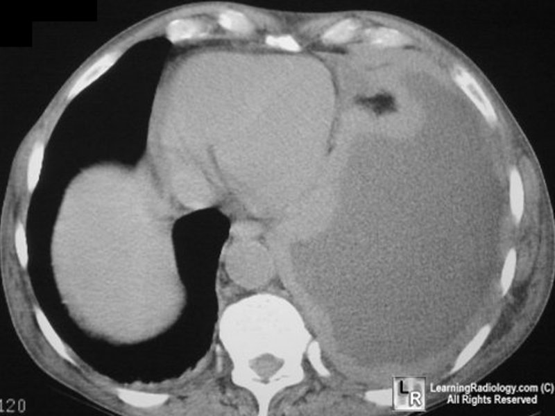
Thick rind of irregular, nodular, malignant
mesothelioma encases the left lung.
There is a large pleural effusion present.
- Extension into interlobar fissures (40-86%)
- Rib destruction in 20% (in advanced disease)
- Ascites (peritoneum involved in 35%)
- CT
- Pleural thickening (92%)
- Thickening of interlobar fissure (86%)
- Pleural effusion (74%)
- Contraction of affected hemithorax (42%):
- Ipsilateral mediastinal shift
- Narrowed intercostal spaces
- Elevation of ipsilateral hemidiaphragm
- Calcified pleural plaques (20%)
- MR (best modality to determine resectability)
- Minimally hyperintense relative to muscle on
T1WI
- Moderately hyperintense relative to muscle on
T2WI
- Metastases to:
- Ipsilateral lung (60%)
- Hilar and mediastinal nodes
- Contralateral lung and pleura (rare)
- Extension through chest wall and diaphragm
- Prognosis
- 10% of occupationally exposed individuals die of
mesothelioma (in 50% pleural + in 50% peritoneal mesothelioma)
- Mean survival time of 5-11 months
- DDx
- Pleural fibrosis from infection (TB, fungal,
actinomycosis)
- Fibrothorax
- Empyema
- Metastatic adenocarcinoma
- Diagnosis
- Video-assisted thoracoscopic surgery (postprocedural radiation therapy of all entry ports for tumor seeding of needle track
[21%])
|
|
|
