|
|
Ventricular Aneurysm
Congenital left ventricular aneurysms
- Rare
- Young black adults
- Produce abnormal bulge in region of left atrium or
- Cardiac enlargement from aortic insufficiency
- Acquired left ventricular aneurysms
- Usually complication of myocardial infarction
- May be associated with
- Persistent congestive heart failure
- Arrhythmias
- Peripheral embolization
- Usually are either true or false aneurysms
True ventricular aneurysms
- Localized outpouching of ventricular cavity
- Associated with dyskinesia
- Wide-mouthed in connection with the LV
- Anterolateral or apical wall
- Frequently not visible on chest x-ray but may produce localized bulge of left heart border
- Paradoxical expansion during systole
- May have rim of calcium in fibrotic wall
- Ventriculography is diagnostic
- Complications
- Thrombus with embolization
- Rarely rupture
- False or pseudoaneurysms
- False aneurysm occurs when left ventricle ruptures into pericardial sac
- Pericardial adhesions contain rupture
- Usually occurs on posterolateral wall
- Diameter of mouth is smaller
- Causes
- Myocardial infarction
- Trauma
- Increase in size over serial films suggests false aneurysm
- This finding indicates the patient requires immediate attention
- High risk of delayed rupture
- Calcification of left ventricular wall takes several years after myocardial infarction
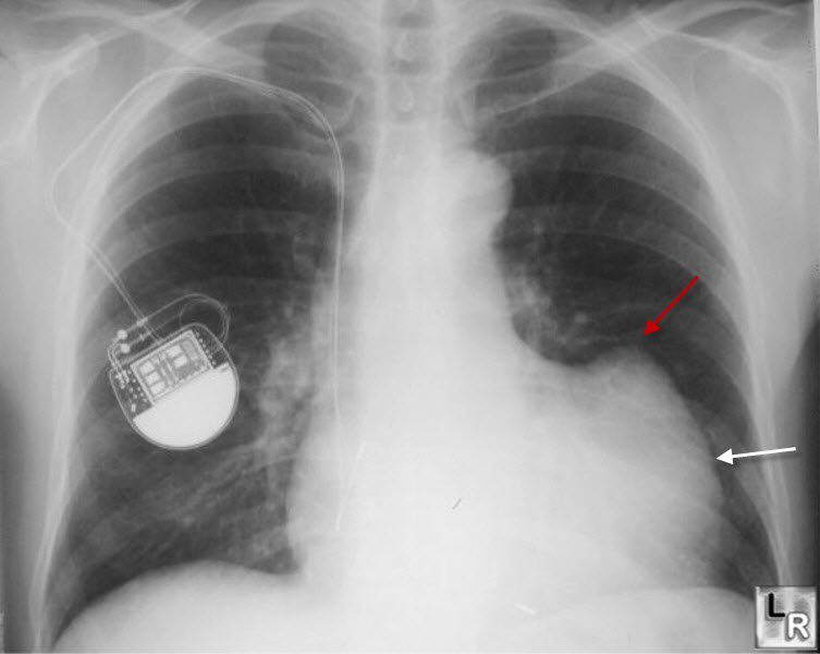
Left Ventricular Aneurysm. There is an unusual contour of the left heart border (white arrow) and an abnormal protuberance in the region of the left ventricle (red arrow).
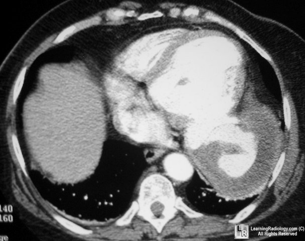
Enhanced CT scan at level of heart shows
very large left ventricular aneurysm partially filled with clot
Cooley and
Schreiber-Radiology of the Heart and Great Vessels-3rd Edition
|
|
|
