|
|
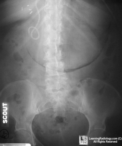
Gastric Emphysema and Emphysematous Gastritis
Gastric Emphysema
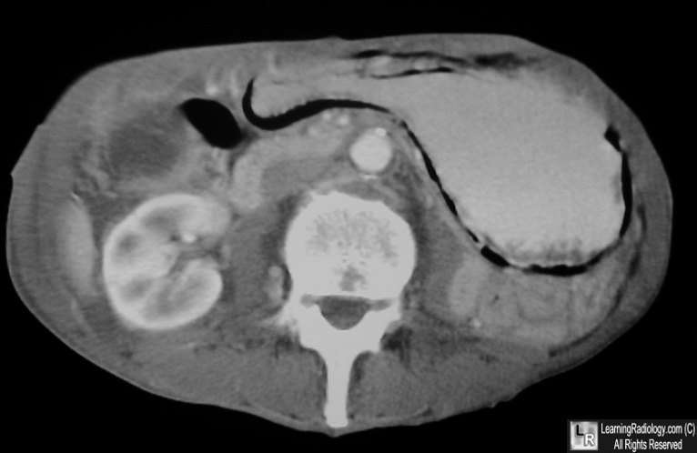
Air outlines the wall of the stomach on an axial scan of the upper abdomen.
Emphysematous gastritis
- Rare and severe gastritis secondary to mucosal
disruption and gas-forming bacterial invasion
- Characterized by air in the wall of the stomach
- Causes:
- Ingestion of toxic material such as corrosives
- Alcohol ingestion
- Trauma
- Gastric infarction
- Ulcer disease
- Submucosa is invaded by gas-forming organisms
which include:
- Hemolytic strep
- Clostridia Welchi
- E. Coli
- Staph aureus
- Clinical:
- Sudden and violent onset of bloody
emesis
- Fever
- Nausea
- Chills
- Leukocytosis
- Imaging
- Linear small gas bubbles in gastric wall
- Gastric emphysema is more linear,
streak-like
- Gas in portal vein
- Prognosis:
- Best way to differentiate emphysematous
gastritis from gastric emphysema:
- Look at patient
- Patients with gastric emphysema are a
asymptomatic from the bowel gas air
- Patients with emphysematous gastritis are
usually deathly ill
|
|
|