|
|
Fahr Disease
Submitted by Andrew Hwang, MD
- Part of a spectrum of diseases characterized by extensive deposition of calcium within the basal ganglia.
- Prominent calcifications may also occur within the dentate nuclei, centrum semiovale, and subcortical white matter.
- Other causes of bilateral basal ganglia calcification included in the differential diagnosis include postinflammatory causes, such as TB, toxoplasmosis, cysticercosis, congenital HIV.
- Clinical presentation is variable.
- Patients may present with progressive dementia, neuromuscular impairment, and dysarthria.
- Parkinsonian symptoms may also occur.
- The disease can present sporadically, but is most often purported to be familial, with variable inheritance patterns.
- There is no known cure, and therapy is supportive in nature.
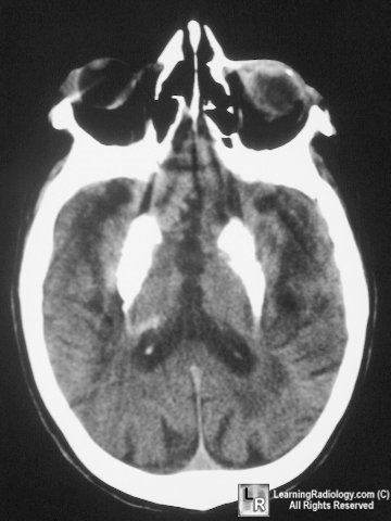 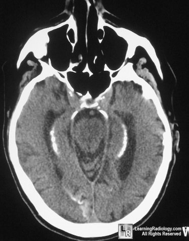
Fahr Disease. Unenhanced CT reveals dense calcifications within the basal ganglia, subcortical white matter of the posterior parietal lobes, and the dentate nuclei of the cerebellum.
Osborn A. Diagnostic Neuroradiology. Mosby, St. Louis; 1994:745-746
Haaga JR. Computed Tomography and Magnetic Resonance Imaging of
the Whole Body. Mosby, St. Louis; 1994
|
|
|
